
Файл:Tubular adenoma high mag.jpg

Алдын ала көрүүнүн көлөмү: 800 × 600 пиксел Башка уруксаттар: 320 × 240 пиксел | 640 × 480 пиксел | 1 024 × 768 пиксел | 1 280 × 960 пиксел | 2 048 × 1 536 пиксел.
Түп нуска файл (2 048 × 1 536 пиксель, файлдын көлөмү: 540 KB, MIME түрү: image/jpeg)
Жыйынтыгы
| Сыпаттама |
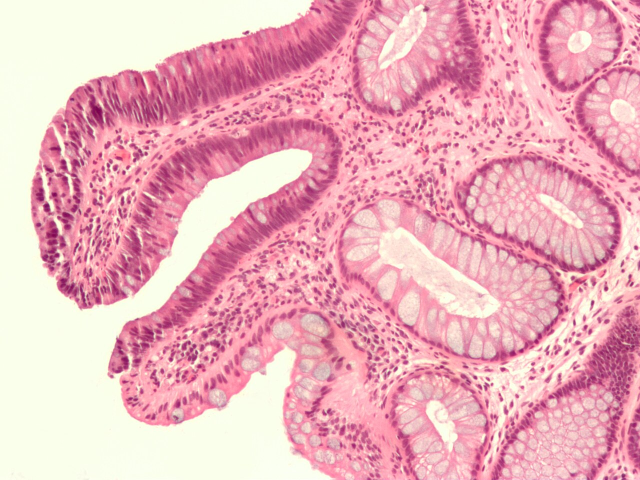
English: Micrograph of a colorectal tubular adenoma. No high grade dysplasia. H&E stain. Specimen removed during a colonoscopy.
The lesional tissue, i.e. dysplastic epithelium, is seen on the left of the image and characterized by nuclear hyperchromatism (i.e. dark purple nuclei), nuclear crowding (i.e. nuclei are bunched-up), elliptical/cigar-shaped nuclei and loss of goblet cells/reduction in the number of goblet cells. Normal colonic type epithelium is seen on the right of the image and characterized by small round nuclei and abundant goblet cells. See also: Related imagesAnother case:
|
| Дата | |
| Булак | Жумушубуз |
| Автор | Nephron |


Лицензиялоо
I, the copyright holder of this work, hereby publish it under the following licenses:
This file is licensed under the Creative Commons Attribution-Share Alike 3.0 Unported license.
- Сиз буларга эркинсиз:
- бөлүшүү – чыгарманы көчүрүү, жайылтуу жана өткөрүп берүү
- ремикс кылуу – чыгарманы ылайыкташтыруу
- Төмөнкү шарттарда:
- атрибуция – Сиз тийиштүү насыя берип, лицензияга шилтеме калтырып жана өзгөртүүлөр болсо көрсөтүшүңүз керек. Сиз муну кандайдыр бир акылга сыярлык жол менен жасай аласыз, бирок лицензиар сизди же сиздин колдонууңузду жактырган кандайдыр бир жол менен эмес.
- Бирдей шарттар боюнча бөлүшүү – If you remix, transform, or build upon the material, you must distribute your contributions under the same or compatible license as the original.

|
Permission is granted to copy, distribute and/or modify this document under the terms of the GNU Free Documentation License, Version 1.2 or any later version published by the Free Software Foundation; with no Invariant Sections, no Front-Cover Texts, and no Back-Cover Texts. A copy of the license is included in the section entitled GNU Free Documentation License. |
Сиз тандаган лицензияны тандай аласыз.
Файлдын тарыхы
Файлдын белгилүү бир учурдагы көрүнүшүн көрүү үчүн тийиштүү убакыт/датаны басыңыз
| Убакыт/дата | Миниатюра | Өлчөм | Колдонуучу | Түшүндүрмө | |
|---|---|---|---|---|---|
| учурдагы | 03:48, 13 февраль 2009 | | 2 048 × 1 536 (540 KB) | Nephron | {{Information |Description={{en|1=Micrograph of a colorectal tubular adenoma. No high grade dysplasia. H&E Stain. Specimen removed during a colonoscopy. The lesional tissue, i.e. dysplastic epithelium, is seen on the left of the image and characterized b |
Шилтемелер
Бул файл төмөнкү баракта колдонулат:
Файлдын глобалдык колдонулушу
Бул файл төмөнкү викилерде колдонулат:
- be.wikipedia.org сайтындагы колдонулушу
- en.wikipedia.org сайтындагы колдонулушу
- hy.wikipedia.org сайтындагы колдонулушу
- kk.wikipedia.org сайтындагы колдонулушу
- pt.wikipedia.org сайтындагы колдонулушу
- ru.wikipedia.org сайтындагы колдонулушу
- te.wikipedia.org сайтындагы колдонулушу
- tg.wikipedia.org сайтындагы колдонулушу
- vi.wikipedia.org сайтындагы колдонулушу